SEM & EDS
Our scanning electron microscope coupled with x-ray analysis (EDS) is used to examine microscopic details of surfaces and sectioned samples, to identify and analyse features of interest for a wide range of investigations:
- metallurgical fractures and failure analysis
- corrosion problems and evaluation of test coupons
- surface coating evaluation and measurement
- polymer and rubber analysis/identification
- textiles and fibres
- glass fragments/contaminants or foreign bodies
- dust particulate identification
We have expertise in analysing samples for a wide range of sectors including:
- Automotive; powertrain, valvetrain, brakes, suspension
- Industrial; lifting equipment, hydraulics, catering
- Oil/Gas/Power Generation
- Medical Devices
- Railway
- Domestic; plumbing fittings and pipe, electrical
- Marine
- Electronic Devices
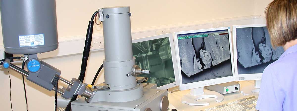
The LEO 435VP is a high-performance, variable pressure scanning electron microscope with a resolution of 4.0nm.
Its 5 axis computer controlled stage is mounted in a specimen chamber measuring 300 x 265 x 190mm and can accommodate specimens weighing up to 0.5kg. Standard automated features include focus, stigmator, gun saturation, gun alignment, contrast and brightness.
The Oxford Isis 300 EDS system is coupled to a thin-window detector for simultaneous analysis of elements boron to uranium. The system has both qualitative and quantitative x-ray analysis capabilities, X-ray map acquisition, high-resolution digital imaging and image analysis.
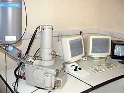
Leo 435VP Scanning Electon Microscope
with Oxford Isis 300 EDS
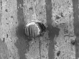
Secondary electron image
of embedded debris in plain bearing
Specification
Scanning Electron Microscope
| Resolution | 4.0nm (High vacuum mode) |
| Accelerating voltage | 0.3 to 30 kV |
| Magnification | x15 to x300,000 |
| Filament | Tungsten hairpin filament |
| Objective lens apertures | 2 position, micrometer controls for precise alignment |
| Specimen chamber | Dimensions: 300mm (long) x 265mm (wide) x 190mm (deep) |
| Specimen stage | 5 axis computer controlled eucentric goniometer x=100mm, y=125mm, z=34mm tilt= 0 to 90°, rotation=360° (continuous) |
| Maximum specimen weight | 0.5kg |
Standards
| ASTM E766-14e1 | Standard Practice for Calibrating the Magnification of a Scanning Electron Microscope |
| ASTM E986-04(2017) | Standard Practice for Scanning Electron Microscope Beam Size Characterization |
| ASTM E1508-12a | Standard Guide for Quantitative Analysis by Energy-Dispersive Spectroscopy |
| ASTM E1829-14 | Standard Guide for Handling Specimens Prior to Surface Analysis |
